
Page 5 - Vulvar Cancer Guidelines Summary fxd
P. 5
DIAGNOSIS AND REFERRAL
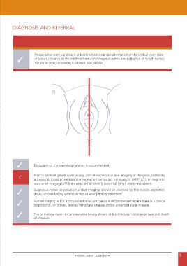
Preoperative work-up should at least include clear documentation of the clinical exam (size
✓ of lesion, distance to the midline/clitoris/anus/vagina/urethra and palpation of lymph nodes).
Picture or clinical drawing is advised (see below).
R
✓ Evaluation of the cervix/vagina/anus is recommended.
C Prior to sentinel lymph node biopsy, clinical examination and imaging of the groin, (either by
ultrasound, (positron emission tomography-) computed tomography ((PET-) CT), or magnetic
resonance imaging (MRI)) are required to identify potential lymph node metastases.
✓ Suspicious nodes (at palpation and/or imaging) should be analysed by fine-needle aspiration
(FNA), or core biopsy when this would alter primary treatment.
✓ Further staging with CT thorax/abdomen and pelvis is recommended where there is a clinical
suspicion of, or proven, (nodal) metastatic disease and/or advanced stage disease.
✓ The pathology report on preoperative biopsy should at least include histological type and depth
of invasion.
• VULVAR CANCER - GUIDELINES • 5