
Page 29 - Ovarian Cancer Surgery - Quality Indicators
P. 29
5.8 QI 8 - Minimum required elements in operative reports
5.8.1 Description of the QI
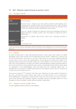
TYPE Process indicator.
DESCRIPTION Operative report is structured. Size and location of disease at the beginning of the
operation must be described. All the areas of the abdominal cavity1) must be described.
If applicable, the size and location of residual disease at the end of the operation, and the
reasons for not achieving complete cytoreduction must be reported.
SPECIFICATIONS Numerator: number of patients with advanced ovarian cancer undergoing cytoreductive
surgery who have a complete operative report that contains all required elements as
defined above.
Denominator: all patients with advanced ovarian cancer undergoing cytoreductive
surgery.
TARGETS 90%.
SCORING RULE 3 if the target is met.
1) ovaries, tubes, uterus, pelvic peritoneum, paracolic gutters, anterior parietal peritoneum, mesentery, peritoneal surface of the colon and bowel, liver, spleen,
greated and lesser omentum, porta hepatis, stomach, Morrison pouch, lesser sac, undersurface of both hemidiaphragms, pelvic and aortic nodes and if applicable
pleural cavity.
5.8.2 Rationale
In another pathology, there is evidence that standardized operative reports result in more complete and reliably
interpretable operative data compared with non-standardized operative reports272. Furthermore, compliance with
the standardized operative report improves over time. In the absence of international validated standardized
operative report in ovarian cancer, some required elements must be reported. Size and location of disease at the
beginning of the operation must be described. All the areas of the abdominal cavity must be described ovaries,
tubes, uterus, pelvic peritoneum, paracolic gutters, anterior parietal peritoneum, mesentery, peritoneal surface of
the colon and bowel, liver, spleen, greated and lesser omentum, porta hepatis, stomach, Morrison pouch, lesser
sac, undersurface of both hemidiaphragms, pelvic and aortic nodes and if applicable pleural cavity . If
applicable, the size and location of residual disease at the end of the operation, and the reasons for not achieving
complete cytoreduction must be reported.
Three previous initiatives26,53,64 published a QI for this topic. Furthermore, the only guideline125 identified for
this topic recommends that operative reports sould include some required elements e xtent of initial disease
before debulking pelvis, midabdomen, or upper abdomen cutoffs: pelvic brim to lower ribs; amount of residual
disease in the same areas after debulking; complete or incomplete resection; if incomplete, indicate the size of
the major lesion and total number of lesions. Indicate if miliary or small lesions.
5.8.3 Summary of available scientific evidence
No directly applicable clinical studies have been identified.
OVARIAN CANCER SURGERY - QUALITY INDICATORS
29